RICK REA: Helping You Grow Through Online Marketing
  • Home
  • Blog
    • Social Media News
    • SEO Marketing News
    • Digital Trends News
    • Photography News
    • Mobile Marketing
    • Business News
    • Gadget News
    • Printing News
  • Contact
  • About
  • Subscribe

Printing News

What Does the Future Hold for Additive Manufacturing for Medical Devices in 2020?

9/4/2019

0 Comments


What Does the Future Hold for Additive Manufacturing for Medical Devices in 2020?

https://ift.tt/2ZxD3f7

Personalised medical devices are often cited to create the greatest opportunity in additive manufacturing (AM). There are a number of notable advantages that AM can bring to enhance the...

View the entire article via our website.


Printing

via 3DPrint.com | The Voice of 3D Printing / Additive Manufacturing https://3dprint.com

September 4, 2019 at 08:54AM
0 Comments

Price Performance Potential Closing the Gap in 3D Printing

9/4/2019

1 Comment


Price, Performance, Potential – Closing the Gap in 3D Printing

https://ift.tt/2LlUBCt

MakerBot, a global leader in the 3D printing industry, can be seen within the rapid prototyping processes of several industry powerhouses, such as Lockheed Martin and KUKA Robotics. Recently,...

View the entire article via our website.


Printing

via 3DPrint.com | The Voice of 3D Printing / Additive Manufacturing https://3dprint.com

September 4, 2019 at 06:39AM
1 Comment

Sharing Knowledge With CELLINKs Ambassador Program

9/4/2019

0 Comments


Sharing Knowledge With CELLINK’s Ambassador Program

https://ift.tt/2zRxs4d

Creating a sharing ecosystem for research projects in bioprinting is key to making scientific findings reproducible and enabling fellow scientists and engineers to contribute to the evergrowing...

View the entire article via our website.


Printing

via 3DPrint.com | The Voice of 3D Printing / Additive Manufacturing https://3dprint.com

September 4, 2019 at 04:09AM
0 Comments

Allevi Bioprint Pro Software Just Released Provides Users with Step-by-Step Bioprinting

9/4/2019

0 Comments


Allevi Bioprint Pro Software Just Released, Provides Users with Step-by-Step Bioprinting

https://ift.tt/32rwZSo

Headquartered in Philadelphia and founded in 2014, the Allevi Bioprint team has spent years in research and development regarding bioprinting, seeking the best results for creating a machine,...

View the entire article via our website.


Printing

via 3DPrint.com | The Voice of 3D Printing / Additive Manufacturing https://3dprint.com

September 4, 2019 at 02:54AM
0 Comments

Titomic Signs Agreement & MoU with GE Additive Company AP&C for Titanium 3D Printing Powder

9/4/2019

0 Comments

 
https://ift.tt/2HK7DY1

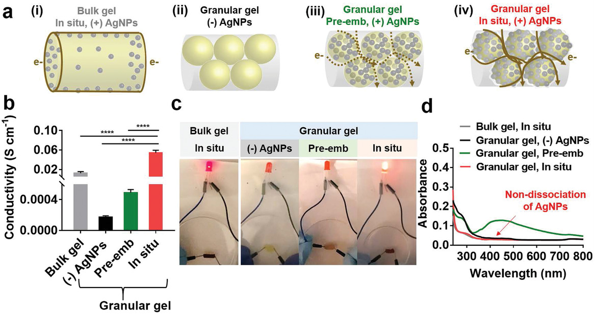
Titomic Signs Agreement & MoU with GE Additive Company AP&C for Titanium 3D Printing Powder

https://ift.tt/2Usb7U7

It hardly seems possible that it’s now been two years since Australian metal 3D printing company Titomic unveiled its patented, innovative Titomic Kinetic Fusion (TKF) process, which is adapted...

View the entire article via our website.


Printing

via 3DPrint.com | The Voice of 3D Printing / Additive Manufacturing https://3dprint.com

September 4, 2019 at 02:51AM
0 Comments

Digital Metal Releases Two New Superalloys for Metal 3D Printing in Extreme Environments

9/4/2019

0 Comments


Digital Metal Releases Two New Superalloys for Metal 3D Printing in Extreme Environments

https://ift.tt/34nTdqr

A few years ago, metal powder producer the Höganäs Group acquired Digital Metal, a small Sweden-based company with a proprietary binder jetting technology of the same name that was developed in...

View the entire article via our website.


Printing

via 3DPrint.com | The Voice of 3D Printing / Additive Manufacturing https://3dprint.com

September 4, 2019 at 02:51AM
0 Comments

Bioprinting at University of Pennsylvania: Impacts on Conductivity in Granular Hydrogels

9/3/2019

0 Comments


Bioprinting at University of Pennsylvania: Impacts on Conductivity in Granular Hydrogels

https://ift.tt/2LiBQjh

To reach the goal of 3D printing human organs, bioprinting must continue to evolve. Researchers are not only aware of this, but as they are part of the process in seeking to make huge impacts in the medical realm, they continue to refine bioprinting in new studies like the one outlined in the recently published ‘Injectable and Conductive Granular Hydrogels for 3D Printing and Electroactive Tissue Support.’

Authors Mikyung Shin, Kwang Hoon Song, Justin C. Burrell, D. Kacy Cullen, and Jason A. Burdick explain their approach to creating form injectable granular hydrogels through the following steps:

  • Hydrogel microparticles are created with water-in-oil emulsions.
  • Microgels are customized during an in situ reduction process.
  • Microgels are ‘jammed’ into extrusion from a syringe.

Conductivity of the hydrogels was easily modified in the new concept created by the authors, based on the assembly of hyaluronic acid microparticles into solids containing metal-phenolic networks. Reduction is based on gallol moieties, common polyphenols with a natural base.

“The gallol moiety has benzene‐1,2,3‐triols, capable of being oxidized to form galloquinones and to donate two electrons per one molecule,” stated the researchers. “When coupled with this oxidation of gallol, metal ions (e.g., M+) are reduced to generate metal nanoparticles (e.g., M0). Furthermore, gallols may act as chelators to form coordinated networks with metal nanoparticles.”

“Further, the intrinsic injectability of granular hydrogels allows fabrication of 3D printed electroactive patterns (e.g., wearable and flexible electronic devices) and electrophysiological support for biological tissues (e.g., myocardium, skeletal muscles).”

The in situ technique offers even greater potential than embedding, refining both conductivity and mechanical properties. The electrical conductivity of the hydrogels was explored further as the authors expected further enhancements due to a large surface area conducive to ‘continuous electric flow.’

Hydrogel structures and their variances impacted conductivity; for instance, within the microgels lacking AgNPs altogether, limited conductivity existed. With the addition of AgNPs, however, conductivity was improved. Size and morphology were noted to make a difference in conductivity too but thought to be dependent on ‘magnitude.’

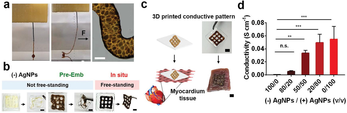
3D printing of conductive hydrogels. a) Images of 3D printing process of the conductive granular hydrogels and morphology of the printed filament. The black arrow indicates a physical force (F) applied to the filament with needle translation during printing, showing self‐supporting of the filament. b) Printability of the granular hydrogels fabricated from microgels without AgNPs (“(−)AgNPs”) or with pre‐embedded (“pre‐Emb”) or in situ synthesized (“in situ”) AgNPs on the polymeric film and their free‐standing stability when removed with forceps. c) Schematic and images of transferring of the printed lattice of the conductive microgels onto porcine myocardium. d) Conductivity of extruded filaments as a function of volumetric mixing ratio v/v of the “(−)AgNPs” or “in situ, (+)AgNPs” microgels. One‐way ANOVA, Dunnett’s test for multiple comparisons to “100/0” filament, n.s. for not significant, **p < 0.01, ***p < 0.001. Scale bars: 100 µm for (a) and 3 mm for (b,c).

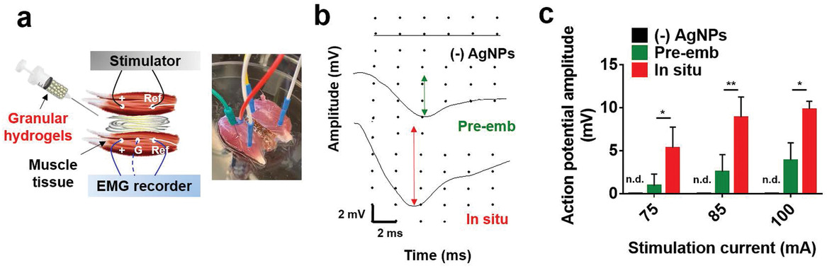
Bridging of conductive tissues with granular hydrogels. a) Schematic and representative image of ex vivo electrical tissue conduction test using isolated skeletal muscles and injection of granular hydrogels between the muscles. b) Electromyogram signals detected in the unstimulated muscle at a stimulation current of 100 mA. The arrows indicate action potential amplitude. c) Action potential amplitude detected in the unstimulated muscle during tissue bridging with the granular hydrogels, from (−)AgNPs, in situ, or pre‐emb microgels. n.d. for not detectable, *p < 0.05, **p < 0.01, two‐way ANOVA.

“Since the metal–phenolic coordination network involved in microgel interactions is dynamic and reversible, applications should consider the potential for dissociation of the microgels and whether further secondary crosslinking is needed for hydrogel stability. For instance, conductivity decreased from 0.05 to 0.01 S cm−1 when incubated for 5 days at body temperature due to a gradual loss of the physicochemical network between microgels,” concluded the researchers.

“The conductive granular hydrogels allowed 3D printable extrusion, fabricating free‐standable constructs on the polymeric film with conductivity as a function of the volumetric ratio of the microgels with/without metal nanoparticles. In addition, the conductive microgels restored electrical conduction by bridging two separated muscle tissues. Our findings present a new technique in the design of soft conductive materials that are also injectable, a promising approach for enhancing electrical conductivity for numerous biomedical applications.”

Bioprinting has led to the use of many different hydrogels—and a range of different research projects around the globe—from shape-shifting hydrogels to those that are created from chitosan or alginate. What do you think of this news? Let us know your thoughts; join the discussion of this and other 3D printing topics at 3DPrintBoard.com.

[Source / Images: ‘

Injectable and Conductive Granular Hydrogels for 3D Printing and Electroactive Tissue Support

’]

Please enable JavaScript to view the comments powered by Disqus.


Printing

via 3DPrint.com | The Voice of 3D Printing / Additive Manufacturing https://3dprint.com

September 3, 2019 at 04:51AM
0 Comments

Royal HaskoningDHV DSM and CEAD Wish to Build Plastic 3D Printed Pedestrian Bridge

9/3/2019

0 Comments


Royal HaskoningDHV, DSM and CEAD Wish to Build Plastic 3D Printed Pedestrian Bridge

https://ift.tt/32lecbt

Engineering company Royal HaskoningDHV is collaborating with polymer company DSM and composites 3D Printing company CEAD to build a pedestrian bridge. CEAD is a Delft based company that is commercializing a large scale continuous fiber 3D printing process. Their CFAM Prime process can make glass or carbon fiber reinforced parts that are four by 2 by 1.5 meters in size. Carbon Fiber Reinforced Polymer materials have a wide range of applications. The design freedom of 3D printing combined with these materials could lead to an entirely new way of constructing bridges and other construction parts. 

We’ve heard a lot about concrete printing over the past years. At the same time, large scale polymer printers have been used for molds for formwork which lets one make large scale parts for tunnels and other structures. CEAD and BAAM systems have experimented with structural parts directly but this is to be the first pedestrian bridge 3D printed out of polymers.

VIDEO

By combining polymers with continuous fiber a lightweight stiff structure can emerge with very high strength. Some issues may remain in some cases such a structure may be too brittle for applications as bridges. In the past, many 3D printed buildings have tended to fail when they come into contact with the elements especially things like freezing temperatures. These things will have to be ironed out but this is a very exciting development indeed.

Maurice Kardas, Business Development Manager at Royal HaskoningDHV, said, 

“This collaboration will bring about a paradigm shift in the way we think of the form and functionality in bridges. Fiber Reinforced Plastic bridges have been known for their long life spans and lower overall costs in comparison with steel bridges. Now we will be using a new 3D printing technology which lets us at scale make fiber-reinforced plastic parts.”

“Through adding sensors to the bridge we can make a ‘digital twin’ of the bridge itself. These sensors can predict and optimize maintenance, ensure safety and lengthen the life span of bridges.”

Patric Duis, Segment Leader Additive Manufacturing bij DSM stated, 

“Using Arnite has significant advantages in bridge building. Instead of traditional materials such as steel and concrete, we can make more environmentally friendly bridges with more flexible design and through using recyclable materials. Designs that would before have been challenging or even impossible..can now be made through 3d printing.” 

Concrete is a huge pollutant and it would be a great development to see bridges made of recyclable materials or bridges that can be recycled. Arnite is a stiff PBT or PET material that could, especially in its PET form be ideal for this. Composites themselves are very difficult to recycle, however.

VIDEO

The team will have to show that these complex materials can also actually be turned into something useful end of life. Many recycling processes for these materials have yet to be developed. Smart manufacturers are already turning to natural fibers to create composites that enable easy end of life recycling.

VIDEO
What’s also interesting is that CEAD has a printing method Fused Granulate Fabrication that works based on granulate. Many of the large scale 3D printing technologies are granulate based since this reduces the cost of parts. Will granulate become a trend in medium-format 3D printing as well, or will these larger printers stick to filament? Will someone develop an integrated recycling solution capable of producing polymer formwork or structures such as bridges out of recycled materials? That, of course, will really turn this into an ideal solution for long-lasting environmentally friendly construction.

Please enable JavaScript to view the comments powered by Disqus.


Printing

via 3DPrint.com | The Voice of 3D Printing / Additive Manufacturing https://3dprint.com

September 3, 2019 at 03:45AM
0 Comments

Interview with Seok-Hwan You of Rokit Healthcare on Bioprinting

9/3/2019

0 Comments


Interview with Seok-Hwan You of Rokit Healthcare on Bioprinting

https://ift.tt/2MSk8Vy

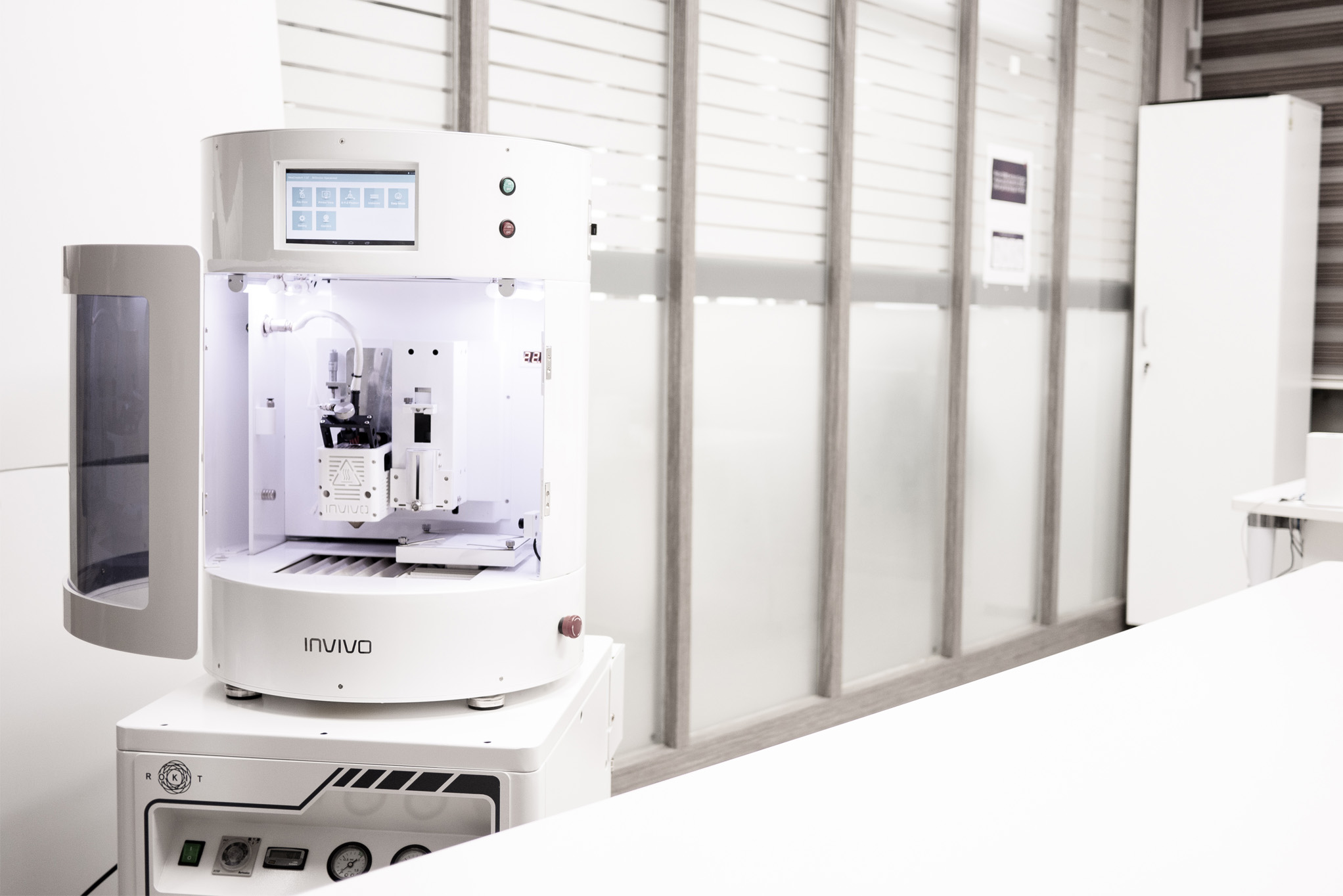
When Seok-Hwan You founded Rokit Healthcare the company was one of the first worldwide to be able to 3D print PEEK and other high-performance materials. It quickly grew to dominate its local Korean medical and bioprinting market before reaching overseas for expansion. Recently the firm pivoted from just selling 3D printers and materials towards offering integrated solutions. With a renewed focus on regenerative healthcare, the firm is offering complete solutions for bioprinting. Rokit Healthcare now offers bioinks, the firm has a tissue bank, a 3D printing service and training. Rokit Healthcare is now furthering its goal to lead in bioprinting. I was very impressed by Rokit’s facilities and staff when I visited the firm. We interviewed Rokit Healthcare CEO Seok-Hwan You to find out more about his vision on bioprinting and goals for the pioneering company.

What is Rokit Healthcare?

ROKIT Healthcare strives to improve the quality of life and health around the world by addressing the problem of aging and age-related diseases with total, healthcare solutions. 3D biofabrication and the development of patient-specific tissue and organ regeneration therapies are our core capabilities. However, we are also involved in the provision of other healthcare programs, such as genetic testing of individuals, customized insurance services, and global medical tours.

Why did you pivot towards regenerative medicine and away from bioprinting?

We have not pivoted “away from bioprinting” per se. It is very much our core scientific technology; it sets the base for personalized therapy solutions we expect to introduce to global hospitals, from patient-specific skin and cartilage regeneration to heart and retina patch biofabrication solutions. However, as previously mentioned, we believe bioprinting must converge with other preventive medicine and diagnostic technologies, digitalization and healthcare management strategies to be truly effective at the level of patient outcomes.
So, we seek to address regenerative medicine and healthcare from a much wider vantage point, with bioprinting as an important but not the only one area of our endeavors.

Why should 3D Print partner with you? 

What sets ROKIT Healthcare apart is that we offer services and insights from a total regenerative medicine solution provider’s perspective rather than a 3D bioprinting device, biomaterial, or 3D printed tissue products company.
As much as 3D bioprinting sits at the center of an exponential tech convergence, a group that can approach the field from various vantage points of health business and economics is likely to be an ideal partner for 3D Print in reaching out to its diverse professional client bases. ROKIT Healthcare is such a group.

What customers are you looking for?

Currently, our priority lies in developing customer bases for our 3D bioprinter and biomaterial platforms. Our focus customers include research groups from universities, government institutes, hospital labs, and pharmaceutical companies. The fields of application range from tissue engineering and regenerative medicine to micro-tissue development for pharmaceutical testing as an alternative to animal experiments. Soon, however, as we introduce 3D bioprinting-based therapy solutions like skin and cartilage regeneration platforms, we expect our client bases to expand beyond life sciences research to doctors and medical device companies.

What is your company culture like?

Like all great companies, we value integrity, excellence, respect, collaboration, and autonomy. But the four key values we as a company live by are: 1) ownership: we encourage strong ownership and autonomous decision-making by employees; 2) detail-orientation: we approach every task with a practice of thoroughly and concisely reviewing product or service execution; 3) no surrender: we do not give up easily and find value in even the littlest 1% possibility against 99% objections; 4) back to the basics: we stick to simplicity and adherence to fundamental principles and values of integrity, discipline, and respect.

At the convergence of these four values stands one key action principle: “Keep Blitz and Simple”. We, ROKIT Healthcare, are committed to maximizing employee freedom to fight the “python of process”. We give unusual amounts of freedom and information to all employees, sharing documents and business plans internally broadly and systematically, because we believe that highly-informed and autonomous employees are capable of good judgment to lead the company to success.

What do you hope to achieve over the next five years?

We envision integrating the 3D bioprinter and its applications into the traditional healthcare services, making the idea of bioprinting as a medical device a reality. Already we have begun the journey this month, with the start of our first clinical study of bioprinting-based skin regeneration for diabetic foot ulcer patients in India (August, 2019). 

Why is it important to bioprint inside the operating theatre?

Bioprinting right by the bedside inside the operating theatre means minimized time, risks, and costs in the transfer of patient cells to the bioprinter and in the transfer of printed tissues back to the patient. It is a new kind of point-of-care personalized healthcare solution that maximizes the benefits of autologous regenerative medicine technologies.

What bioprinting materials are you excited about?

Nowadays, there are many kinds of bioinks in the market, including synthetic and natural polymers. But, they are not all applicable to the human body. We say “Aging is a Disease; Nature is the Best Therapy”. In that sense, we are excited about the whole extracellular matrix (ECM)-based bioinks that are derived from the ‘Human Body’. We believe the ECM is Nature.

What new developments are very interesting to you?

When it comes to traditional diagnostics of cancer, regardless of its kinds, doctors have been treating their patients only with bulk RNA-based analysis. But, as many of us realize today in the cancer research field, we know that the reason cancer is so hard to treat is that it is an extremely heterogeneous population of cells. We understand that each cell is its own universe. Understanding each of these universes is only possible by single-cell RNA analysis, and this will be key to taking any step closer to finding effective treatments for cancer. The scRNA technology may have much room to mature, but we’re working on it excited about its potential.

What products do you have?

We supply INVIVO, our signature 3D bioprinter. Plus, we supply 3D printers for material engineering and advanced prototyping in biomedical fields with materials like PEEK and ULTEM.

Do you have high hopes for PEEK? PCL? Other materials?

We have been paying a great deal of attention to broadening applications for medical-grade PCL and PEEK, especially in the applications of bone regenerative matrix and customized pill fabrication. However, the greatest focus of our energy lies not on synthetic plastics, but on natural ingredients like human ECM as a supportive materials for 3D printed living cells. 

What are the challenges in bioprinting?

The biggest challenge that all industrial players of 3D bioprinting face is closing the gap between the technology we supply and the actual needs of our customers in the bioprinting research. A key part of these needs is to understand that bio 3D printing, unlike industrial 3D printing, is not only about manufacturing structures with architectural stability but about promoting cell ingrowth and considering the impact of manufacture on cell viability. Based on a constant probe into such understanding, we are building a base for developing next-generation bioprinting technologies and biomaterial applications.

Please enable JavaScript to view the comments powered by Disqus.


Printing

via 3DPrint.com | The Voice of 3D Printing / Additive Manufacturing https://3dprint.com

September 3, 2019 at 02:57AM
0 Comments

Investigating 3D Printed Biomodels in Experimental Blood Flow Studies

9/3/2019

0 Comments


Investigating 3D Printed Biomodels in Experimental Blood Flow Studies

https://ift.tt/2MRvTLU

There are many applications for 3D printing in the biomedical research community, such as lab-on-a-chip tools, surgical planning, and drug delivery. Yet another is 3D biomodels, which is the focus of a study, titled “Low Cost 3D printed biomodels for biofluid mechanics applications,” published by a group of Portuguese researchers from the University of Minho and the Polytechnic Institute of Bragança. Carlos L. Faria, Diana Pinho, Jorge Santos, Luís M. Gonçalves, and Rui Lima discussed the fabrication of 3D biomodels for use in hemodynamic (relating to the flow of blood within the body’s organs and tissues) experimental flow studies.

The abstract states, “This paper shows the ability of the desktop 3D printers (also known as low cost 3D printers) to produce 3D biomodels able to be used on hemodynamic experimental flow studies. Overall, this paper shows that Fused Deposition Modelling (FDM) process combined with polydimethylsiloxane (PDMS) replication molding is a promising way to produce affordable biomedical devices to perform hemodynamic studies at both macro and micro scale levels.”

Biomodels are devices – physical or virtual – that replicate the form or geometry of a biological structure, like an artery. They can be used to perform in vitro and numerical experiments, and for their paper, the researchers presented an overview of successful polydimethylsiloxane (PDMS) biomodels made on desktop 3D printers, combined with PDMS replication molding, in order to complete in vitro blood flow studies at the microscale  and macroscale levels.

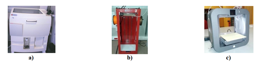
Fig. 1 3D printers used to make macro models: (a) Zprinter 310 Plus (b) Big Builder and (c) Cube 3D

The team tested three different 3D printers to perform micro and macro flow studies. Macro models of human carotid arteries were initially built on the Zprinter 310 Plus, but the team later switched to the extrusion-based systems of the Big Builder and Cube 3D.

“The intracranial aneurysm model and the 3D models of the micro devices were fabricated by depositing the thermoplastic material acrylonitrile butadiene styrene (ABS) on a stage layer-by-layer trough an extrusion nozzle,” the researchers wrote. “The advantages of this method are the low cost, the speed, and the ability to apply different kinds of thermoplastic materials, such as the ABS, polylactic acid (PLA), and nylon.”

CT (TC) scans were used to 3D print (TDP) the human carotid artery geometry for the PDMS models in the macro flow studies. Scan IP software was used to segment the TC images, and the file was converted to an STL. The 3D printed models were then put inside a molding box, and the biocompatible PDMS was poured over the master mold and cured. After it cooled, the model was removed from the mold box “where the inlet and outlet tubes were connected.”

“For the case of the PDMS microfluidic devices (dimensions from 5 mm down to 0.3 mm) to perform flow studies at micro scale level, the 3D models were obtained by the FDM process combined by PDMS replication molding,” the team explained. “The PDMS (Sylgard® 186, Dow Corning) was prepared also by mixing the prepolymer with the curing agent at 10:1 ratio and poured onto the printed models placed in the bottom of a petri dish and cured in an oven at 80 °C for 20 minutes. In parallel, another mixture of PDMS (20:1 ratio) was spin coated at 5000 rpm for 2 minutes (VTC-100 Vacuum Spin Coater) over a glass slide and cured in an oven at 80 °C for 20 minutes. By using a blade, the micro channels were cut off and the inlet/outlet holes of the fluid were done by using a fluid-dispensing tip. Finally, the channels were sealed by using the coated glass slide. Note that, to achieve a strong adhesion of the materials, the device was placed inside the oven at 80 °C for 24 h.”

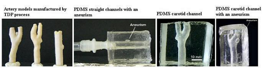
Fig. 2 3D printed models and PDMS transparent flow channels

The team then presented an overview of work by other researchers focused on using PDMS 3D models for macro flow studies, such as 3D printed carotid artery models with and without aneurysms and a wall expansion assessment of an FDM 3D printed intracranial aneurysm model.

“The PDMS transparent models obtained from the 3D models fabricated by the TDP technique have shown to be a promising way to perform in vitro blood flow studies through anatomically realistic replica of a human carotid artery with and without aneurisms,” they explained about the former study.

“A promising approach to understand the mechanical behavior of aneurysms is by measuring the deformability of the walls by means of optical techniques,” they continued about the latter study.

“Pinho et al. [16] have developed a methodology able to measure experimentally the displacement field of an in vitro intracranial aneurysm model. This method is a combination of aneurysm in vitro models fabricated in PDMS…and the use of an Electronic Speckle Pattern Interferometry (ESPI) technique.”

This second study showed that wall thickness is important in terms of initiating aneurysm growth and later rupture, and that 3D printing can help validate numerical simulations of aneurysms, and find more details about what causes ruptures.

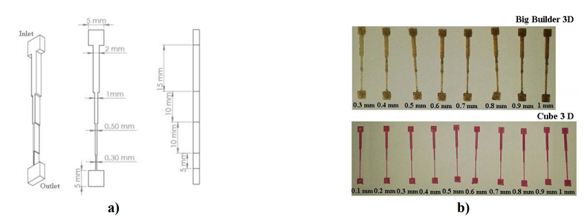
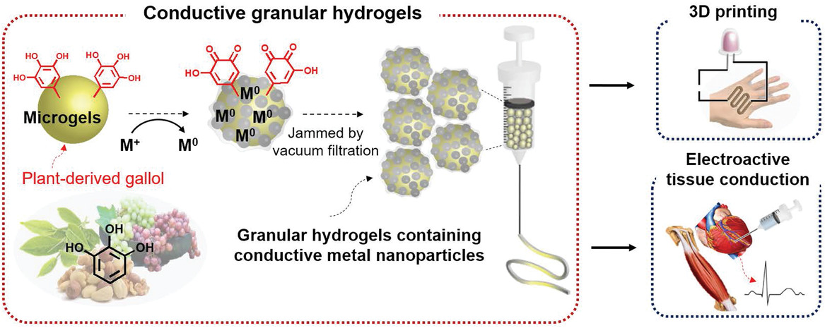
Fig. 5 (a) Model drawn in the Solidworks CAD software and dimensions tested in this study, and (b) microdevice master models 3D printed in the Big Builder and Cube 3D printers

The researchers also discussed several experimental in vitro micro blood flow studies, using 3D printed microdevices, that have taken place to provide a better understanding of the blood flow phenomena in microvessels and biomedical microdevices. They noted that a soft lithography method with expensive equipment is the most common way to make microfluidic devices, which is why it’s “important to explore low cost fabrication techniques.”

“The desktop 3D printers have shown potential to fabricate channels around 1mm, however few works have explored the ability of the low cost 3D printers to fabricate microfluidic devices,” they wrote. “In the present work we have tested the fabrication of several microchannels down to 0.3 mm by using two different desktop printers based on the FDM process.”

They 3D printed several ABS master models of microchannels to perform in vitro blood flow studies, and also made PDMS flow devices from the 3D printed master molds to investigate the cell-free layer (CFL) blood flow phenomenon that occurs during micro circulation.

“From the results obtained from all the tested PDMS flow devices we have only observed a clear CFL at the PDMS devices fabricated by the Cube 3D printer with a height of 0.1 mm. Fig.6 shows clearly that at a height of 0.1 mm there is a tendency to generate a cell depleted region around the wall of the microchannel,” the researchers wrote. “In contrast, this tendency was not observed for the heights of 0.5 and 1 mm. Although, these results have demonstrated that is possible to have the formation of a CFL within the microchannels produced by the Cube 3D printer, the width of the flow channels need to be further reduced in order to obtain a cross section with a geometry more close to real microvessels.”

Fig. 6 In vitro blood flow visualizations at PDMS microfluidic devices fabricated by the Cube 3D printer

The team is pleased with the “extremely encouraging” results they got from the 3D printed devices combined with PDMS molds.

“We believe that this combination is a promising technique to perform more realistic in vitro blood studies through anatomical models and consequently improve our current understanding of the origin and development of cardiovascular diseases,” they concluded.

Discuss this and other 3D printing topics at 3DPrintBoard.com or share your thoughts below.

Please enable JavaScript to view the comments powered by Disqus.


Printing

via 3DPrint.com | The Voice of 3D Printing / Additive Manufacturing https://3dprint.com

September 3, 2019 at 02:51AM
0 Comments
<<Previous

    Categories

    All
    3-D Printed Ultrasound Models
    3-D Printing
    3D Printing
    Tools For The Visually Impared
    Visually Impared

    Archives

    May 2023
    April 2023
    March 2023
    February 2023
    January 2023
    December 2022
    November 2022
    October 2022
    September 2022
    August 2022
    July 2022
    June 2022
    May 2022
    April 2022
    March 2022
    February 2022
    January 2022
    December 2021
    November 2021
    October 2021
    September 2021
    August 2021
    July 2021
    June 2021
    May 2021
    April 2021
    March 2021
    February 2021
    January 2021
    December 2020
    November 2020
    October 2020
    September 2020
    September 2019
    August 2019
    July 2019
    June 2019
    May 2019
    April 2019
    March 2019
    February 2019
    January 2019
    December 2018
    November 2018
    October 2018
    September 2018
    August 2018
    July 2018
    June 2018
    May 2018
    April 2018
    March 2018
    February 2018
    January 2018
    December 2017
    November 2017
    October 2017
    September 2017
    August 2017
    July 2017
    June 2017
    May 2017
    April 2017
    March 2017
    February 2017

    RSS Feed

Powered by Create your own unique website with customizable templates.
  • Home
  • Blog
    • Social Media News
    • SEO Marketing News
    • Digital Trends News
    • Photography News
    • Mobile Marketing
    • Business News
    • Gadget News
    • Printing News
  • Contact
  • About
  • Subscribe