
|
Investigating 3D Printed Biomodels in Experimental Blood Flow Studies https://ift.tt/2MRvTLU There are many applications for 3D printing in the biomedical research community, such as lab-on-a-chip tools, surgical planning, and drug delivery. Yet another is 3D biomodels, which is the focus of a study, titled “Low Cost 3D printed biomodels for biofluid mechanics applications,” published by a group of Portuguese researchers from the University of Minho and the Polytechnic Institute of Bragança. Carlos L. Faria, Diana Pinho, Jorge Santos, Luís M. Gonçalves, and Rui Lima discussed the fabrication of 3D biomodels for use in hemodynamic (relating to the flow of blood within the body’s organs and tissues) experimental flow studies.
Biomodels are devices – physical or virtual – that replicate the form or geometry of a biological structure, like an artery. They can be used to perform in vitro and numerical experiments, and for their paper, the researchers presented an overview of successful polydimethylsiloxane (PDMS) biomodels made on desktop 3D printers, combined with PDMS replication molding, in order to complete in vitro blood flow studies at the microscale and macroscale levels. 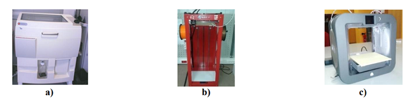
Fig. 1 3D printers used to make macro models: (a) Zprinter 310 Plus (b) Big Builder and (c) Cube 3D The team tested three different 3D printers to perform micro and macro flow studies. Macro models of human carotid arteries were initially built on the Zprinter 310 Plus, but the team later switched to the extrusion-based systems of the Big Builder and Cube 3D.
CT (TC) scans were used to 3D print (TDP) the human carotid artery geometry for the PDMS models in the macro flow studies. Scan IP software was used to segment the TC images, and the file was converted to an STL. The 3D printed models were then put inside a molding box, and the biocompatible PDMS was poured over the master mold and cured. After it cooled, the model was removed from the mold box “where the inlet and outlet tubes were connected.”
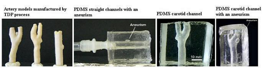
Fig. 2 3D printed models and PDMS transparent flow channels The team then presented an overview of work by other researchers focused on using PDMS 3D models for macro flow studies, such as 3D printed carotid artery models with and without aneurysms and a wall expansion assessment of an FDM 3D printed intracranial aneurysm model.
This second study showed that wall thickness is important in terms of initiating aneurysm growth and later rupture, and that 3D printing can help validate numerical simulations of aneurysms, and find more details about what causes ruptures. 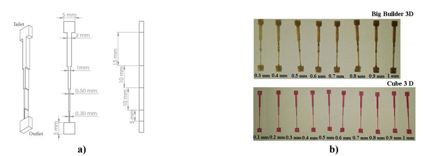
Fig. 5 (a) Model drawn in the Solidworks CAD software and dimensions tested in this study, and (b) microdevice master models 3D printed in the Big Builder and Cube 3D printers The researchers also discussed several experimental in vitro micro blood flow studies, using 3D printed microdevices, that have taken place to provide a better understanding of the blood flow phenomena in microvessels and biomedical microdevices. They noted that a soft lithography method with expensive equipment is the most common way to make microfluidic devices, which is why it’s “important to explore low cost fabrication techniques.”
They 3D printed several ABS master models of microchannels to perform in vitro blood flow studies, and also made PDMS flow devices from the 3D printed master molds to investigate the cell-free layer (CFL) blood flow phenomenon that occurs during micro circulation.

Fig. 6 In vitro blood flow visualizations at PDMS microfluidic devices fabricated by the Cube 3D printer The team is pleased with the “extremely encouraging” results they got from the 3D printed devices combined with PDMS molds.
Discuss this and other 3D printing topics at 3DPrintBoard.com or share your thoughts below. Please enable JavaScript to view the comments powered by Disqus.Printing via 3DPrint.com | The Voice of 3D Printing / Additive Manufacturing https://3dprint.com September 3, 2019 at 02:51AM
0 Comments
Leave a Reply. |
Categories
All
Archives
April 2023
|
 RSS Feed
