
|
Bioprinting at University of Pennsylvania: Impacts on Conductivity in Granular Hydrogels https://ift.tt/2LiBQjh To reach the goal of 3D printing human organs, bioprinting must continue to evolve. Researchers are not only aware of this, but as they are part of the process in seeking to make huge impacts in the medical realm, they continue to refine bioprinting in new studies like the one outlined in the recently published ‘Injectable and Conductive Granular Hydrogels for 3D Printing and Electroactive Tissue Support.’
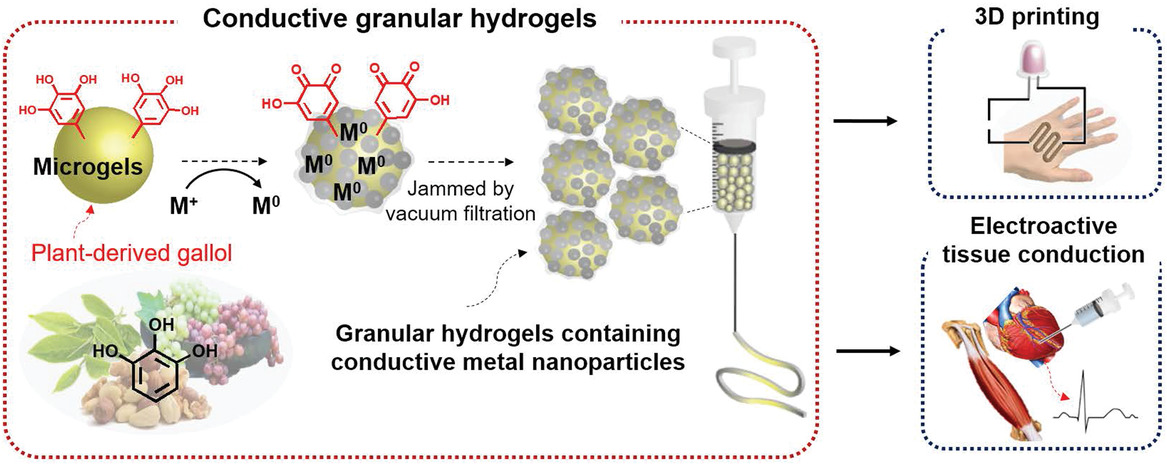
Conductivity of the hydrogels was easily modified in the new concept created by the authors, based on the assembly of hyaluronic acid microparticles into solids containing metal-phenolic networks. Reduction is based on gallol moieties, common polyphenols with a natural base.
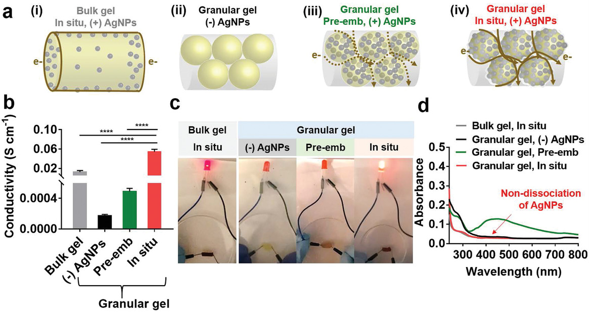
The in situ technique offers even greater potential than embedding, refining both conductivity and mechanical properties. The electrical conductivity of the hydrogels was explored further as the authors expected further enhancements due to a large surface area conducive to ‘continuous electric flow.’
Hydrogel structures and their variances impacted conductivity; for instance, within the microgels lacking AgNPs altogether, limited conductivity existed. With the addition of AgNPs, however, conductivity was improved. Size and morphology were noted to make a difference in conductivity too but thought to be dependent on ‘magnitude.’ 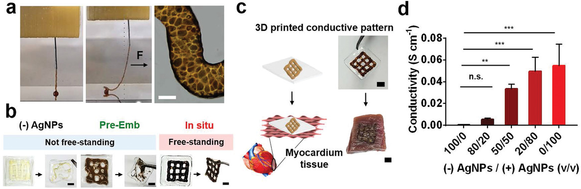
3D printing of conductive hydrogels. a) Images of 3D printing process of the conductive granular hydrogels and morphology of the printed filament. The black arrow indicates a physical force (F) applied to the filament with needle translation during printing, showing self‐supporting of the filament. b) Printability of the granular hydrogels fabricated from microgels without AgNPs (“(−)AgNPs”) or with pre‐embedded (“pre‐Emb”) or in situ synthesized (“in situ”) AgNPs on the polymeric film and their free‐standing stability when removed with forceps. c) Schematic and images of transferring of the printed lattice of the conductive microgels onto porcine myocardium. d) Conductivity of extruded filaments as a function of volumetric mixing ratio v/v of the “(−)AgNPs” or “in situ, (+)AgNPs” microgels. One‐way ANOVA, Dunnett’s test for multiple comparisons to “100/0” filament, n.s. for not significant, **p < 0.01, ***p < 0.001. Scale bars: 100 µm for (a) and 3 mm for (b,c). 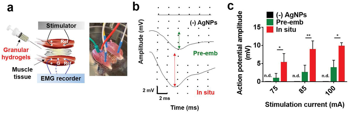
Bridging of conductive tissues with granular hydrogels. a) Schematic and representative image of ex vivo electrical tissue conduction test using isolated skeletal muscles and injection of granular hydrogels between the muscles. b) Electromyogram signals detected in the unstimulated muscle at a stimulation current of 100 mA. The arrows indicate action potential amplitude. c) Action potential amplitude detected in the unstimulated muscle during tissue bridging with the granular hydrogels, from (−)AgNPs, in situ, or pre‐emb microgels. n.d. for not detectable, *p < 0.05, **p < 0.01, two‐way ANOVA.
Bioprinting has led to the use of many different hydrogels—and a range of different research projects around the globe—from shape-shifting hydrogels to those that are created from chitosan or alginate. What do you think of this news? Let us know your thoughts; join the discussion of this and other 3D printing topics at 3DPrintBoard.com. [Source / Images: ‘ Injectable and Conductive Granular Hydrogels for 3D Printing and Electroactive Tissue Support’] Please enable JavaScript to view the comments powered by Disqus.Printing via 3DPrint.com | The Voice of 3D Printing / Additive Manufacturing https://3dprint.com September 3, 2019 at 04:51AM
0 Comments
Leave a Reply. |
Categories
All
Archives
April 2023
|
 RSS Feed
