
Mayo Clinic Researchers 3D Printing Models & Surgical Guides for Chest Wall Reconstructive Surgery6/27/2019 Mayo Clinic Researchers 3D Printing Models & Surgical Guides for Chest Wall Reconstructive Surgery https://ift.tt/2FCcF85
The researchers 3D printed two different models during the case study, with one demonstrating anterior fractured cartilage, and the right side acting as a mirror for the ‘normal ribs’ to demonstrate misalignment of the other side. The models were used to plan the surgery, involving a thoracotomy incision and more. Chest trauma often requires surgical stabilization of rib fracture (SSRF). 3D printing has been reported as useful during intricate cases in the past for creating chest wall prostheses, but not with reconstruction using standard surgical tools.
The patient studied during this research had actually been stabbed, causing the trauma to his left chest area—also resulting in cardiac arrest. Surgery was performed, but the pulmonary hernia developed later and was repaired in a second procedure. He continued to be in pain, however, especially when laughing or coughing or stretching. As surgeons prepared to restore his chest wall with plates and bicortical screws, they used 3D printed models consisting of:
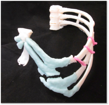
Model photos demonstrate non-union at the costal cartilage. The models offered a clear perspective for the surgeons regarding areas like the disruption at the costosternal joint which was causing left chest distortion. As they continued to study the 3D printed models, the doctors were able to decide on their surgical approach. The authors point out, however, that reconstruction of the chest wall is a complicated procedure, ‘fraught with difficulties,’ to include:
With 3D modeling, however, surgeons can optimize the entire surgical and possible implantation process too.
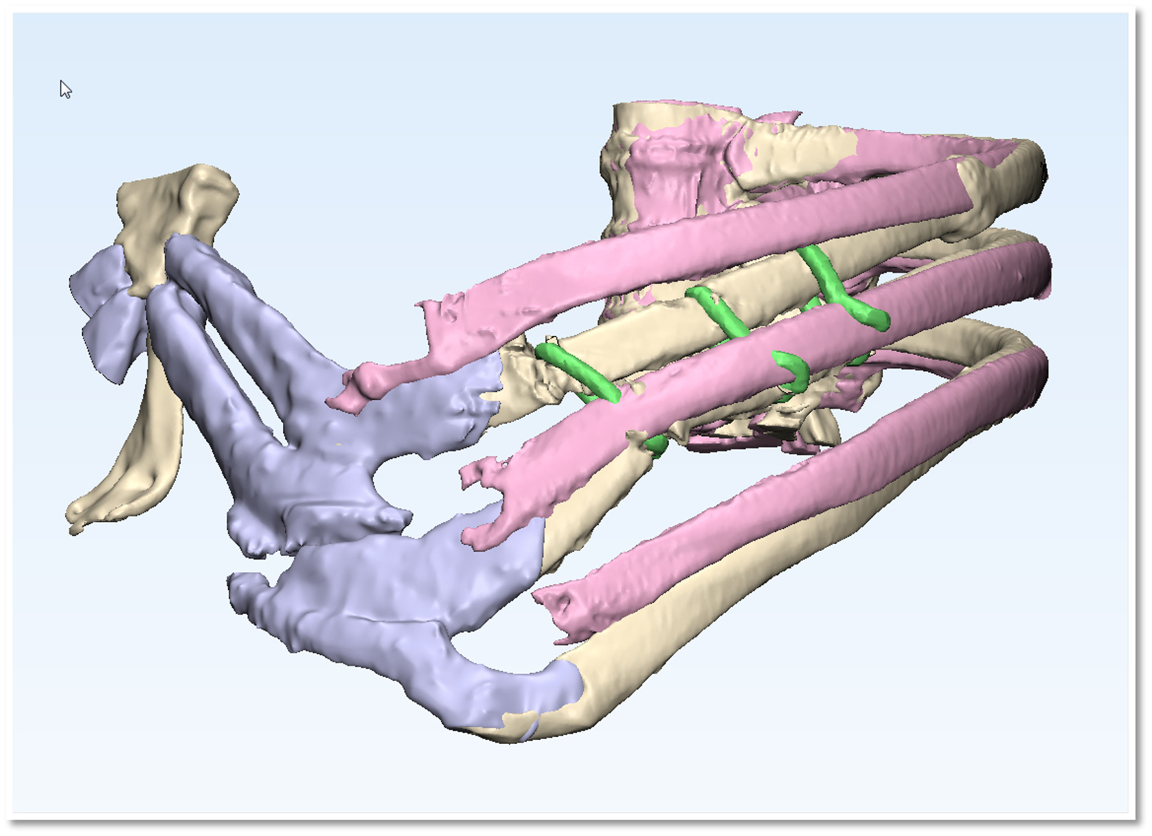
Overlaid model demonstrates the degree of mal-alignment based on mirror image data from the contra-lateral side. As 3D printed models can be printed from the medical facility, all the benefits of 3D printing technology are recognized from affordability in production to speed to customization—and best of all, patient-specific treatment.
3D printing holds a special place in the world of medicine as the technology has made such sweeping changes in the way medical professionals are able to diagnose and treat other medical issues too like brain tumors, examine heart conditions with cardiac patches, and even create devices like 3D printed protheses for children with undeveloped eyes—and countless other issues making life much more tolerable for patients around the world. What do you think of this news? Let us know your thoughts! Join the discussion of this and other 3D printing topics at 3DPrintBoard.com. 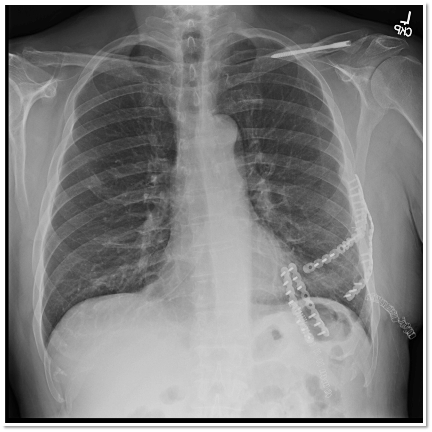
Post-operative film showing reduced and plated ribs and costosternal articulation. [Source / Images: 3D printed modeling contributes to reconstruction of complex chest wall instability] Please enable JavaScript to view the comments powered by Disqus.Printing via 3DPrint.com | The Voice of 3D Printing / Additive Manufacturing https://3dprint.com June 27, 2019 at 04:42AM
0 Comments
Leave a Reply. |
Categories
All
Archives
April 2023
|

 RSS Feed
