
|
https://ift.tt/2GaSr8a
3D Printing Used to Predict Behavior of Replacement Heart Valves https://ift.tt/2rsQEkx
3D printing has been studied by several different institutions as a method for aiding in a process called transcatheter aortic valve replacement, or TAVR. More than one in eight people aged 75 and older in the United States develop moderate to severe blockage of the aortic valve, often caused by calcified deposits that build up on the valve’s leaflets and prevent them from fully opening and closing. Many of these patients are not healthy enough to undergo open heart surgery, so TAVR is an alternative that involves deploying an artificial valve via a catheter inserted into the aorta. Inserting the properly sized valve is critical; if the valve is too small, it can dislodge or leak around the edges, and if it’s too large it can rip through the heart, which can be fatal. It’s a challenge to select the correct size without directly examining the patient’s heart, however. But researchers at the Wyss Institute for Biologically Inspired Engineering at Harvard University have come up with a 3D printing workflow that creates models of individual patients’ aortic valves using CT scan data, in addition to a “sizer” device that helps cardiologists determine the proper valve size. The work is documented in a paper entitled “Pre-procedural fit-testing of TAVR valves using parametric modeling and 3D printing.” The research was carried out in collaboration with researchers and physicians from Brigham and Women’s Hospital, The University of Washington, Massachusetts General Hospital, and the Max Planck Institute of Colloids and Interfaces.
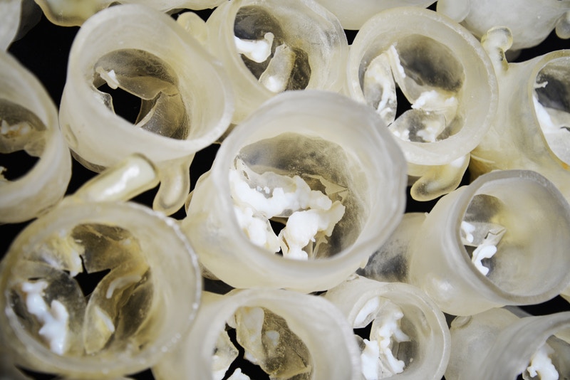
When a patient needs a new heart valve, they typically get a CT scan, but while the outer wall of the aorta and any calcified deposits are easily seen on a scan, the leaflets that open and close the valve are often too thin to show up clearly.
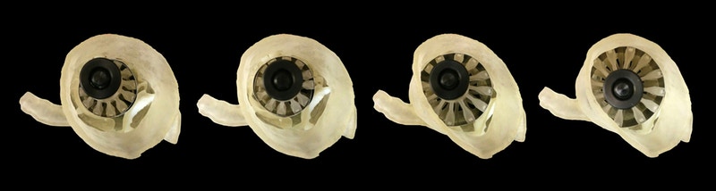
To address this issue, Ahmed Hosny, who was a Research Fellow at the Wyss Institute at the time, created a software program that uses parametric modeling to generate virtual 3D models of the leaflets using seven coordinates on each patient’s valve that are visible on CT scans. The 3D models were then merged with the CT data and adjusted so that they fit into the valve correctly. The resulting model, which incorporates the leaflets and their calcified deposits, was then 3D printed in multiple materials. The researchers also 3D printed a custom sizer device that fits inside the 3D printed valve and expands and contracts to determine what size artificial valve would best fit each patient. They then wrapped the sizer with a thin layer of pressure-sensing film to map the pressure between the sizer and the 3D-printed valves and their associated calcified deposits, while gradually expanding the sizer.
The multi-material 3D printed valve models could also more accurately mimic the behavior of real heart valves during artificial valve deployment, as well as provide haptic feedback as the sizer is expanded. The researchers tested the system against data from 30 patients who had already undergone TAVR procedures. 15 of those patients had developed leaks from too-small valves. The researchers predicted, based on how well the sizer fit into the 3D printed models of their aortic valves, what size valve each patient should have received, and whether they would experience leaks after the procedure. The system successfully predicted leak outcome in 60 to 73% of the patients, depending on the type of valve each patient had received, and determined that 60% of the patients had received the correctly sized valve.
The researchers have made their leaflet modeling software and 3D printing protocol available online for free.
Discuss this and other 3D printing topics at 3DPrintBoard.com or share your thoughts below. [Source/Images: Wyss Institute]
Printing via 3DPrint.com | The Voice of 3D Printing / Additive Manufacturing https://3dprint.com December 10, 2018 at 03:39PM
0 Comments
Leave a Reply. |
Categories
All
Archives
April 2023
|

 RSS Feed
